2026-10-07 ブラウン大学
<関連情報>
- https://www.brown.edu/news/2026-10-07/cell-movement-tracking
- https://pubs.rsc.org/lc/article/doi/10.1039/D6LC00530F/1363585/Label-Free-All-Electrical-Tracking-of-Individual
メガピクセルCMOS容量センサーを用いた、ラベルフリーの全電気式による個々の細胞および集団細胞の移動追跡
Label-Free All-Electrical Tracking of Individual and Collective Cell Migration on a Megapixel CMOS Capacitance Sensor
Hyuntae Jeong;Pushkaraj Joshi;Yinshi Hu;Jiwon Kim;Anh H Vu;Jacob K Rosenstein;Ian Y Wong
Lab on a Chip Published:30 September 2026
DOI:https://doi.org/10.1039/D6LC00530F
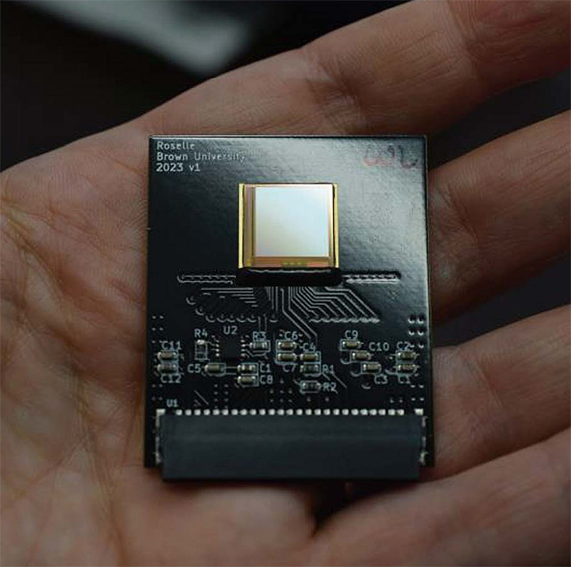
Label-free tracking of adherent cell migration could enable important insights into biological processes such as tissue repair, inflammatory response, or cancer progression. Nevertheless, visualizing unlabeled animal cells using optical microscopy remains challenging due to low contrast as well as frequent changes in cell shape and number. A promising alternative uses electrical capacitance measurements, which are sensitive to cell adhesion to electrode surfaces. However, prior examples often utilized electrodes with areas larger than single cells, resulting in averaged readouts over multiple cells. Here, we demonstrate label-free, live-cell tracking using a capacitance sensor array with more than 1 million pixels on a 10 micron pitch across an area larger than 1 square centimeter. We show that single cell morphology can be clearly segmented, and then used to reconstruct migration and proliferation dynamics using optical flow. We further track the spreading of multicellular spheroids, revealing fast-moving peripheral regions led by a collective leader cell “front.” Finally, we demonstrate label-free imaging of millimeter-scale honeycomb-shaped tissues without the multi-image stitching often required for conventional microscopy. We utilize mutual capacitance measurements with electrically-programmable electrode spacing to reconstruct topographical features of these engineered tissues. Overall, CMOS capacitance imaging arrays enables label-free imaging spanning from single cells to large tissues, in a portable and scalable format for settings where optical microscopy may be difficult to access.