2026-07-09 東北大学
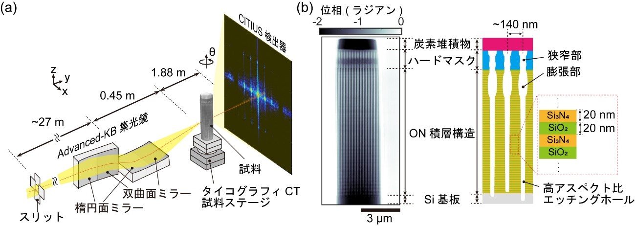
図1. テンダーX線タイコグラフィCT測定系の模式図と再構成像
<関連情報>
- https://www.tohoku.ac.jp/japanese/2026/07/press20260709-02-EtchedHoles.html
- https://pubs.aip.org/aip/apl/article/129/1/011109/3397935/Nondestructive-structural-evaluation-of-high
軟質X線プティコグラフィーとコンピュータ断層撮影による高アスペクト比エッチング孔の非破壊構造評価
Nondestructive structural evaluation of high-aspect-ratio-etched holes by tender x-ray ptychography with computed tomography
Naru Okawa;Nozomu Ishiguro;Yuhei Sasaki;Masaki Abe;Shuntaro Takazawa;Mihiro Ikenaga;Yukio Takahashi
Applied Physics Letters Published:July 08 2026
DOI:https://doi.org/10.1063/5.0339243
Nondestructive evaluation of high-aspect-ratio (HAR) structures in advanced memory devices is essential yet remains challenging at the nanoscale. In this study, we demonstrate tender x-ray ptychography for the characterization of HAR-etched hole structures with depths of approximately 10 μm. Reconstructed two-dimensional projection images, with a spatial resolution of 38.5 nm, reveal features such as necking at the amorphous carbon mask and tapered hole profiles. Furthermore, a three-dimensional (3D) electron density map was reconstructed using ptychographic x-ray computed tomography, achieving a spatial resolution of 67.5 nm. This approach enables direct visualization of etch-rate variations associated with aspect-ratio-dependent etching. These results establish a powerful nondestructive methodology for three-dimensional nanoscale evaluation of HAR structures, with significant potential for applications in advanced memory device development.