(Quantum brain sensors could spot dementia after University of Sussex scientists find they track brain waves)
2021-11-19 英国・サセックス大学

・ サセックス大学、ブライトン・アンド・サセックス・メディカルスクール、ブライトン大学およびドイツ国立計量研究所(PTB)が、高感度の量子脳センサーを開発。
・ 同センサーは、ニューロン(神経細胞)の発火時に生成される磁場を検出し、脳の経時的な変化を測定して脳内で移動する電気信号の速度(バイオマーカー)を追跡。将来的には、認知症、筋萎縮性側索硬化症(ALS)やパーキンソン病等の脳疾患の特定に役立つ可能性がある。
・ 本研究では、空間と時間の双方で極めて正確な結果を提示する量子センサーを初めて実証。脳の信号の場所を特定する有意性は他の研究にてすでに示されているが、量子センサーによる信号の正確なタイミングの特定は今回が初めて。
・ 頭蓋骨に近い場所でのセンシングが可能なため、空間と時間の両分解能が向上し、現在の EEG(脳波計)や fMRI(機能的磁気共鳴画像法)を大幅に超える精度が期待できる。
・ 同量子センサーに含まれるルビジウム原子にレーザー光を照射し、同原子が磁界の変化を感知して異なる光を放出する。この放射光の変動が脳における磁気的活動の変化を提示する。
・ 脳磁図(MEG)技術と量子センサーを組み合わせることで、脳活動の非侵襲的なプローブ技術を実現。脳への送信後に戻ってくる信号を読み取る既存の侵襲的でリスクのある脳スキャン方法とは異なり、MEG は脳内で起こっている現象を外部から受動的に計測する。
・ 現在の MEG 装置は高価で大型のため、臨床診察での利用は困難。量子センサー技術の開発は、研究室の高度に制御された環境から実社会での実用への転換に不可欠なもの。
URL: https://www.sussex.ac.uk/broadcast/read/56791
<NEDO海外技術情報より>
(関連情報)
Scientific Reports 掲載論文(フルテキスト)
Improved spatio-temporal measurements of visually evoked fields using optically-pumped
magnetometers
URL: https://www.nature.com/articles/s41598-021-01854-7
Abstract
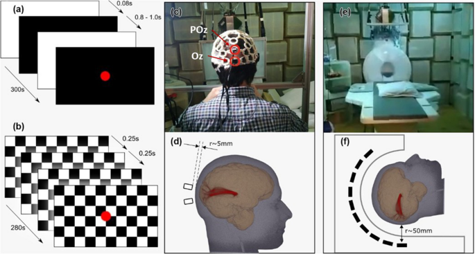
Recent developments in performance and practicality of optically-pumped magnetometers (OPMs) have enabled new capabilities in non-invasive brain function mapping through magnetoencephalography. In particular, the lack of cryogenic operating conditions allows for more flexible placement of sensor heads closer to the brain, leading to improved spatial resolution and source localisation capabilities. Through recording visually evoked brain fields (VEFs), we demonstrate that the closer sensor proximity can be exploited to improve temporal resolution. We use OPMs, and superconducting quantum interference devices (SQUIDs) for reference, to measure brain responses to flash and pattern reversal stimuli. We find highly reproducible signals with consistency across multiple participants, stimulus paradigms and sensor modalities. The temporal resolution advantage of OPMs is manifest in a twofold improvement, compared to SQUIDs. The capability for improved spatio-temporal signal tracing is illustrated by simultaneous vector recordings of VEFs in the primary and associative visual cortex, where a time lag on the order of 10–20 ms is consistently found. This paves the way for further spatio-temporal studies of neurophysiological signal tracking in visual stimulus processing, and other brain responses, with potentially far-reaching consequences for time-critical mapping of functionality in healthy and pathological brains.


