2026-05-27 東北大学
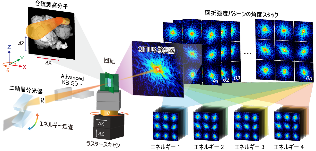
図1. 硫黄K吸収端近傍におけるX線タイコグラフィCT測定の模式図
<関連情報>
- https://www.tohoku.ac.jp/japanese/2026/05/press20260527-01-sulfur.html
- https://www.nature.com/articles/s41598-026-52630-4
硫黄K吸収端プティコグラフィー断層撮影法を用いた、マイクロメートル厚のポリマー中の硫黄化学状態の三次元イメージング Three-dimensional imaging of sulfur chemical states in polymers with micrometer thickness using sulfur K-edge ptychographic tomography
Yuhei Sasaki,Nozomu Ishiguro,Masaki Abe,Shuntaro Takazawa,Naru Okawa,Mihiro Ikenaga,Fusae Kaneko & Yukio Takahashi
Scientific Reports Published:16 May 2026
DOI:https://doi.org/10.1038/s41598-026-52630-4 Unedited version
Abstract
Three-dimensional visualization of chemical bonding states in light-element materials remains challenging, particularly for micrometer-thick specimens, due to strong X-ray absorption and limited photon flux in the tender X-ray regime. Here three-dimensional chemical-state imaging of sulfur in sulfurized poly(n-butyl methacrylate) (SPBMA) samples up to ~ 3.3 µm thick is demonstrated using ptychographic X-ray computed tomography at the sulfur K-edge. A high-resolution ptychographic CT system at the NanoTerasu BL10U beamline enables quantitative mapping of electron density, sulfur concentration, and spectral metrics derived from multi-energy imaging, that serve as proxies for sulfur–sulfur and sulfur–carbon bonding states, with ~ 80 nm spatial resolution. Multi-energy volumetric analysis using four carefully chosen photon energies reveals pronounced spatial heterogeneity in sulfur bonding, distinguishing sulfur–sulfur and sulfur–carbon bonds that are inaccessible from spatially averaged spectra. This study establishes sulfur K-edge ptychographic tomography as a potential platform for three-dimensional chemical-state imaging of light-element materials, enabling new nanoscale investigations of functional polymers and energy-related materials.