2023-01-04 カリフォルニア大学校アーバイン校(UCI)
この発見は、最近Science Advances誌の表紙を飾りましたが、医療やハイテクを含む多くの分野の研究者や産業界が、様々な材料や組織の化学組成を迅速に可視化するのに役立つと期待されています。
「中赤外光は、化学結合に関連しています。」と、カリフォルニア大学化学学部の博士号候補で、この論文の主執筆者であるDave Knez氏は言います。「この技術によって、我々は、より確信を持って、サンプル中に特定の化学物質、または、特定の化学結合が存在すると言うことができます。
この手法の開発の鍵は、画像を構成するのに必要な赤外線波長を素早く捉えて区別できるようにすることでした。これは、スマートフォンのカメラが、可視光領域の異なる色、つまり異なる波長を記録して写真を作成するのと同様である、とカリフォルニア大学レーザー分光研究所のドミトリー・フィッシュマン所長は語っている。Fishman氏によると、化学結合は、スペクトルの赤外線領域でのみ振動し、光を吸収する。これまでは、精細な画像を作ることは容易ではなかった。新しい技術では、「赤外線に “色 “を見ることができるのです。そして、色によって分光線が浮かび上がり、それが化学的な “指紋 “になるのです」。
そのため、「人体組織を含む、見ている物質の組成」を正確に評価することがはるかに容易になります、とUCI化学教授で論文の共著者であるEric Potmaは述べています。医療分野では、病気にかかった組織の分析に最も応用されるだろうと、Potma教授は述べ、この方法はそのような検査を劇的にスピードアップし、改善することができると説明している。また、この技術は、時間の経過に伴う化学組成の変化を検出することもできる。これは、化学反応やプロセスを追跡する際に役立つだろう。
<関連情報>
- https://news.uci.edu/2023/01/04/uc-irvine-scientists-create-new-chemical-imaging-method/
- https://www.science.org/doi/full/10.1126/sciadv.ade4247
- https://opg.optica.org/optica/fulltext.cfm?uri=optica-8-7-995&id=453041
- https://www.nature.com/articles/s41377-020-00369-6
中赤外域の高精細・高速スペクトルイメージング Spectral imaging at high definition and high speed in the mid-infrared
David Knez ,Benjamin W. Toulson,Anabel Chen ,Martin H. Ettenberg ,Hai Nguyen ,Eric O. Potma ,Dmitry A. Fishman
Science Advances Published:16 Nov 2022
DOI: 10.1126/sciadv.ade4247

Abstract
Spectral imaging in the mid-infrared (MIR) range provides simultaneous morphological and chemical information of a wide variety of samples. However, current MIR technologies struggle to produce high-definition images over a broad spectral range at acquisition rates that are compatible with real-time processes. We present a novel spectral imaging technique based on nondegenerate two-photon absorption of temporally chirped optical MIR pulses. This approach avoids complex image processing or reconstruction and enables high-speed acquisition of spectral data cubes (xyω) at high-pixel density in under a second.
中赤外領域における高速化学選択的3Dイメージング Rapid chemically selective 3D imaging in the mid-infrared
Eric O. Potma, David Knez, Yong Chen, Yulia Davydova, Amanda Durkin, Alexander Fast, Mihaela Balu, Brenna Norton-Baker, Rachel W. Martin, Tommaso Baldacchini, and Dmitry A. Fishman
Optica Published: July 7, 2021
DOI:https://doi.org/10.1364/OPTICA.426199
Abstract
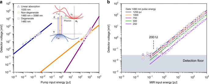
The emerging technique of mid-infrared optical coherence tomography (MIR-OCT) takes advantage of the reduced scattering of MIR light in various materials and devices, enabling tomographic imaging at deeper penetration depths. Because of challenges in MIR detection technology, the image acquisition time is, however, significantly longer than for tomographic imaging methods in the visible/near-infrared. Here we demonstrate an alternative approach to MIR tomography with high-speed imaging capabilities. Through femtosecond nondegenerate two-photon absorption of MIR light in a conventional Si-based CCD camera, we achieve wide-field, high-definition tomographic imaging with chemical selectivity of structured materials and biological samples in mere seconds.
シリコン系カメラにおける非変性2光子吸収を用いた赤外化学イメージング Infrared chemical imaging through non-degenerate two-photon absorption in silicon-based cameras
David Knez,Adam M. Hanninen,Richard C. Prince,Eric O. Potma & Dmitry A. Fishman
Light:Science and Applications Published:20 July 2020
DOI:https://doi.org/10.1038/s41377-020-00369-6

Abstract
Chemical imaging based on mid-infrared (MIR) spectroscopic contrast is an important technique with a myriad of applications, including biomedical imaging and environmental monitoring. Current MIR cameras, however, lack performance and are much less affordable than mature Si-based devices, which operate in the visible and near-infrared regions. Here, we demonstrate fast MIR chemical imaging through non-degenerate two-photon absorption (NTA) in a standard Si-based charge-coupled device (CCD). We show that wide-field MIR images can be obtained at 100 ms exposure times using picosecond pulse energies of only a few femtojoules per pixel through NTA directly on the CCD chip. Because this on-chip approach does not rely on phase matching, it is alignment-free and does not necessitate complex postprocessing of the images. We emphasize the utility of this technique through chemically selective MIR imaging of polymers and biological samples, including MIR videos of moving targets, physical processes and live nematodes.


