2026-07-27 バージニア工科大学(Virginia Tech)

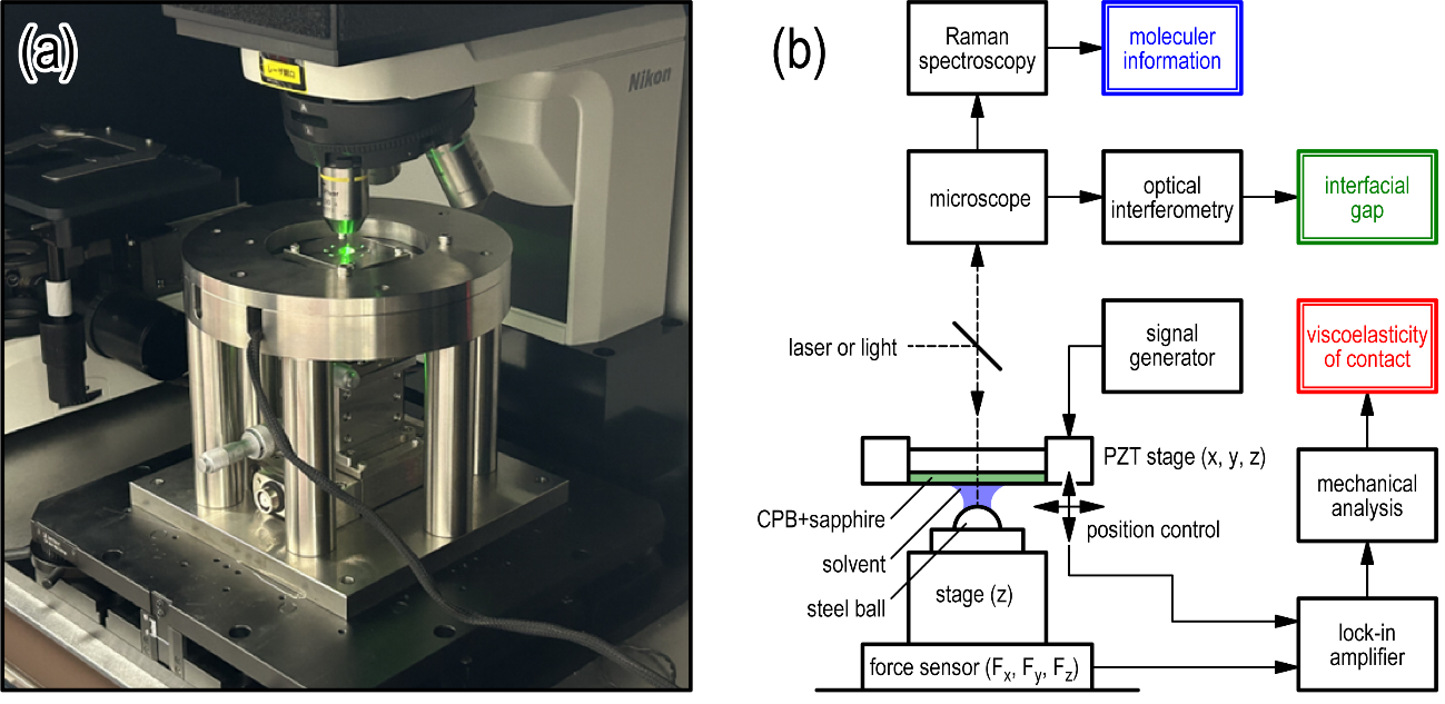
A sensor placed in a wound model with bacterial growth is examined under a microscope linked to an ultrafast laser system used to study sensor regeneration and tissue-relevant environments. Photo courtesy of Aditya Garg.
<関連情報>
- https://news.vt.edu/articles/2026/07/eng-ece-biosensor.html
- https://advanced.onlinelibrary.wiley.com/doi/10.1002/advs.76330
フェムト秒レーザーナノキャビテーションによるSERS活性プラズモンナノギャップの再生と生体界面における縦方向分子センシング Femtosecond-Laser Nanocavitation Regenerates SERS-Active Plasmonic Nanogaps for Longitudinal Molecular Sensing at Biointerfaces
Aditya Garg, Ze Zong, Meitong Nie, Stacie E. Deaver, Elizabeth M. Van Order, Elieser Mejia, Peter Vikesland, Erin S. Gloag, Wei Zhou
Advanced Science Published: 03 July 2026
DOI:https://doi.org/10.1002/advs.76330
ABSTRACT
Longitudinal, long-term molecular monitoring is critical for personalized medicine, yet protein adsorption in biofluids rapidly fouls bio-interfaced sensors and restricts analyte access to sensing regions. Despite advances in antifouling coatings and regeneration strategies, repeated restoration of surface-enhanced Raman spectroscopy (SERS) sensitivity in static, protein-rich media remains difficult without degrading nanogap integrity, limiting longitudinal sensing. This work introduces a regenerative molecular sensor based on multiresonant plasmonic nanoprotruding meshes (MPNMs) that co-localizes SERS sensing and nanocavitation-based actuation within nanogaps anchored on a biocompatible polymeric mesh. The nanogaps are engineered to support an electric-dipole resonance for SERS enhancement and a magnetic-dipole resonance for photothermal conversion, enabling femtosecond-laser-triggered nanocavitation within SERS-active nanogaps. Upon femtosecond-laser irradiation, collapse of vapor nanobubbles (≈200 ns lifetime) generates thermomechanical forces that detach and displace foulants with micron-scale precision to regenerate the nanogaps while preserving nanomorphology and optical performance. In undiluted human serum, regeneration restores detection limit for the Pseudomonas aeruginosa virulence factor pyocyanin from 2.0 µm to a clinically relevant 3.9 nm after 24 h of fouling. Repeated regeneration cycles enable spatiotemporal profiling of dynamic molecular signatures from P. aeruginosa biofilms in wound models over 24 h, establishing a self-regenerating platform for longitudinal molecular monitoring in protein-rich biosystems.