2026-08-20 東京大学,理化学研究所,科学技術振興機構
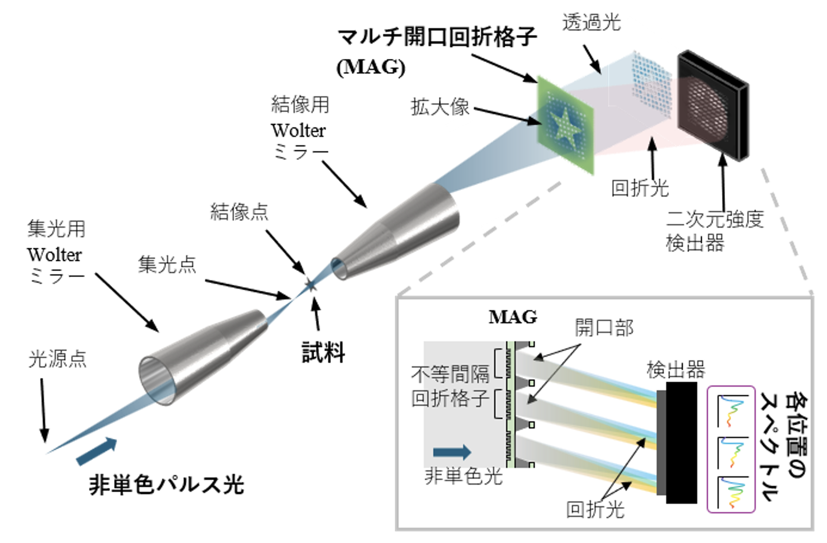
図1:フェムト秒軟X線ハイパースペクトル顕微鏡の原理図
<関連情報>
- https://www.issp.u-tokyo.ac.jp/maincontents/news2.html?pid=31865
- https://opg.optica.org/optica/fulltext.cfm?uri=optica-13-8-1643
単一ショットX線パルスによるフェムト秒ハイパースペクトルイメージング Femtosecond hyperspectral imaging with a single-shot X-ray pulse
Yoko Takeo, Satoru Egawa, Noboru Furuya, Kyota Yoshinaga, Kai Sakurai, Yu Nakata, Shigeki Owada, Takashi Tanaka, Hidekazu Mimura, Makina Yabashi, and Takashi Kimura
Optica Published: August 19, 2026
DOI:https://doi.org/10.1364/OPTICA.599397
Abstract
Single-shot hyperspectral imaging was demonstrated in the soft X-ray region by combining a multi-aperture grating (MAG) with Wolter-mirror microscopy. The MAG simultaneously segments the magnified image into 417 spatial channels and disperses each channel onto a two-dimensional detector. This system functions as a dispersive spectrometer with two-dimensional spatial sampling, obtaining absorption spectra at each point of the magnified image in a single pulse. At SACLA BL1, a field of view of 39µm ×39µm was obtained, and a spatial sampling of 2.4µm(H)×1.6µm (V), defined by the MAG aperture pitch, was achieved. A grating-limited spectral resolving power of E/△E ≈ 103around 100 eV was theoretically estimated, limited by the grating line count per aperture. The system is applied to shot-to-shot measurements of the spatially resolved beam spectrum and to single-shot spectro-microscopy of a silicon nitride thin film near its absorption edge. These results establish a route to chemical-state-sensitive imaging of heterogeneous and potentially non-repeatable samples.