2026-11-12 東北大学
東北大学大学院工学研究科の佐々木雄平大学院生、高橋幸生教授らの研究グループは、3GeV高輝度放射光施設NanoTerasu(ナノテラス)のビームラインBL10Uを利用し、テンダーX線によるX線タイコグラフィで7.4 nmの投影空間分解能を実現した。テンダーX線領域では、従来、10 nmを下回る高分解能イメージングの実現が困難であった。研究では、エネルギー4.5 keVのX線を用い、Advanced Kirkpatrick-Baez全反射集光ミラーと高速積分型X線画像検出器CITIUSを組み合わせ、複数の検出器位置で取得した回折データを統合して画像を再構成した。その結果、金ナノキューブの微細構造を明瞭に可視化し、7.4 nmという高い空間分解能を達成した。本技術は、硫黄やリンなどの吸収端を利用した電池・触媒材料の元素分布や化学状態の高分解能イメージングに加え、半導体デバイス内部の微細構造、界面、欠陥の非破壊観察への応用が期待される。材料の反応・劣化メカニズムの解明や次世代電子デバイスの研究開発に貢献する成果である。
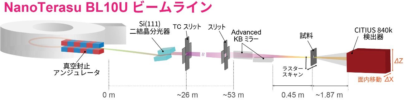
図1. NanoTerasu BL10Uビームラインでのタイコグラフィ測定レイアウト。
<関連情報>
- https://www.tohoku.ac.jp/japanese/2026/11/press20261112-02-NanoTerasu.html
- https://doi.org/10.1107/S1600577526011549
NanoTerasu BL10Uにおける高分解能タイコグラフィーのテンダーX線領域への拡張(Extending high-resolution ptychography into the tender X-ray regime at NanoTerasu BL10U)
Yuhei Sasaki, Nozomu Ishiguro, Masaki Abe, Shuntaro Takazawa, Naru Okawa, Yukio Takahashi
Journal of Synchrotron Radiation 2026-11-05
DOI: 10.1107/S1600577526011549
Tii-summary
本研究では、NanoTerasuのビームラインBL10Uにおいて、テンダーX線領域(2~5 keV)で高分解能タイコグラフィーを実現するための撮像技術を開発した。タイコグラフィーは、試料を走査しながら取得した回折パターンから、レンズを使用せずに試料の構造を高解像度で再構成するX線イメージング手法である。研究グループは、4.5 keVのX線を用い、高性能Kirkpatrick–Baez集光ミラーと高感度CITIUS検出器を組み合わせて実験を実施した。さらに、複数の検出器位置で取得した回折パターンを統合して画像を再構成することで、取得可能な空間周波数範囲を拡張した。金ナノキューブの位相投影像を用いたエッジ応答解析では、約7.4 nmの空間分解能を達成した。本成果は、テンダーX線領域における高分解能観察能力を実証し、元素吸収端を利用した材料・デバイスの微細構造解析や元素選択的イメージングの高度化につながるものである。
This study extends high-resolution X-ray ptychography into the tender X-ray regime using the BL10U beamline at NanoTerasu, a 3 GeV synchrotron radiation facility. Ptychography is a lensless imaging technique that reconstructs sample structures from diffraction patterns collected during scanning. Experiments were performed at 4.5 keV using advanced Kirkpatrick–Baez focusing mirrors and a high-performance CITIUS detector. To extend the accessible spatial-frequency range, diffraction patterns acquired at multiple detector positions were jointly used for image reconstruction. Edge-response analysis of the reconstructed phase projection of gold nanocubes demonstrated a spatial resolution of approximately 7.4 nm. These results establish high-resolution imaging capability in the tender X-ray region and enhance the potential of NanoTerasu BL10U for nanoscale structural characterization, particularly in studies exploiting elemental absorption edges and the complementary phase and absorption contrast of tender X-rays.
This study extends high-resolution X-ray ptychography into the tender X-ray regime using the BL10U beamline at NanoTerasu, a 3 GeV synchrotron radiation facility. Ptychography is a lensless imaging technique that reconstructs sample structures from diffraction patterns collected during scanning. Experiments were performed at 4.5 keV using advanced Kirkpatrick–Baez focusing mirrors and a high-performance CITIUS detector. To extend the accessible spatial-frequency range, diffraction patterns acquired at multiple detector positions were jointly used for image reconstruction. Edge-response analysis of the reconstructed phase projection of gold nanocubes demonstrated a spatial resolution of approximately 7.4 nm. These results establish high-resolution imaging capability in the tender X-ray region and enhance the potential of NanoTerasu BL10U for nanoscale structural characterization, particularly in studies exploiting elemental absorption edges and the complementary phase and absorption contrast of tender X-rays.