2026-11-17 名古屋大学
<関連情報>
- https://www.nagoya-u.ac.jp/researchinfo/result/2026/11/20261117-engg.html
- https://pubs.acs.org/ancac3/article/doi/10.1021/acsnano.6c12991/5464870/Bandgap-and-Interface-Engineering-of-Ag-Au-S
近赤外高輝度発光と深部組織イメージングに向けたAg–Au–S量子ドットのバンドギャップ・界面工学(Bandgap and Interface Engineering of Ag–Au–S Quantum Dots toward Bright Near-Infrared Emission and Deep-Tissue Bioimaging)
Kazutaka Akiyoshi, Hinako Hayashi, Mariko Hasegawa, Eunyeong Ko, Nurmanita Rismaningsih, Chie Miyamae, Hiroyuki Okada, Tatsuya Kameyama, Hiroki Sato, Yusuke Ohshima, Shota Yamada, Hiroshi Yukawa, Yoshinobu Baba, Tsukasa Torimoto
ACS Nano (2026):2026-11-05
DOI:10.1021/acsnano.6c12991
Visual Abstract
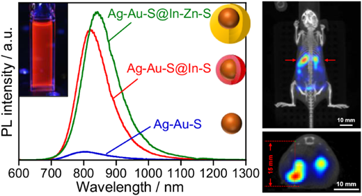
Source: Akiyoshi et al., ACS Nano (2026). https://doi.org/10.1021/acsnano.6c12991. CC BY 4.0.
本研究では、銀(Ag)、金(Au)、硫黄(S)からなる低毒性の多元素半導体量子ドットを開発し、組成制御と異種半導体界面の設計によって近赤外発光特性を改善した。AgとAuの組成比を調整することで、バンドギャップを1.39~1.88 eV、発光ピークを745~1130 nmの範囲で制御できた。さらに、量子ドット表面をIn–Sで被覆すると発光量子収率は3.4%から33.6%に向上し、Znを導入したIn–Zn–S処理では最大50.5%に達した。3-メルカプトプロピオン酸による表面修飾で水溶化した後も、発光量子収率27.0%を維持し、生体関連媒体中で高い発光安定性を示した。細胞毒性試験で低い毒性が確認され、マウスを用いた実験では皮膚表面から15 mm以上の深さにある組織から近赤外発光を検出した。組成と界面の精密制御によって、発光効率、水分散性、安定性を両立した成果であり、生体深部イメージング用プローブへの応用が期待される。
This study developed low-toxicity multicomponent semiconductor quantum dots composed of silver (Ag), gold (Au), and sulfur (S), improving their near-infrared emission through composition control and heterointerface engineering. By adjusting the Ag/Au ratio, the bandgap was tuned from 1.39 to 1.88 eV, while the photoluminescence peak shifted across 745–1130 nm. In–S surface coating increased the photoluminescence quantum yield from 3.4% to 33.6%, and subsequent Zn incorporation into the In–Zn–S surface layer further raised it to as high as 50.5%. After surface modification with 3-mercaptopropionic acid, the quantum dots became water-dispersible while retaining a quantum yield of 27.0% and exhibiting high emission stability in biologically relevant media. Low cytotoxicity was confirmed in cell tests, and in vivo experiments in mice detected near-infrared emission from tissues located more than 15 mm beneath the skin surface. These findings demonstrate that precise control of composition and interfaces can combine high emission efficiency, aqueous dispersibility, and stability, supporting the potential use of these quantum dots as probes for deep-tissue bioimaging.