2024-06-28 カリフォルニア工科大学(Caltech)
<関連情報>
- https://www.caltech.edu/about/news/new-computational-microscopy-technique-provides-more-direct-route-to-crisp-images
- https://www.nature.com/articles/s41467-024-49126-y
- https://www.nature.com/articles/nphoton.2013.187
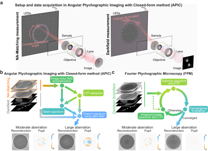
収差補正された閉形式複素場再構成による高解像度、大視野ラベルフリーイメージング High-resolution, large field-of-view label-free imaging via aberration-corrected, closed-form complex field reconstruction
Ruizhi Cao,Cheng Shen & Changhuei Yang
Nature Communications Published:03 June 2024
DOI:https://doi.org/10.1038/s41467-024-49126-y

Abstract
Computational imaging methods empower modern microscopes to produce high-resolution, large field-of-view, aberration-free images. Fourier ptychographic microscopy can increase the space-bandwidth product of conventional microscopy, but its iterative reconstruction methods are prone to parameter selection and tend to fail under excessive aberrations. Spatial Kramers–Kronig methods can analytically reconstruct complex fields, but is limited by aberration or providing extended resolution enhancement. Here, we present APIC, a closed-form method that weds the strengths of both methods while using only NA-matching and darkfield measurements. We establish an analytical phase retrieval framework which demonstrates the feasibility of analytically reconstructing the complex field associated with darkfield measurements. APIC can retrieve complex aberrations of an imaging system with no additional hardware and avoids iterative algorithms, requiring no human-designed convergence metrics while always obtaining a closed-form complex field solution. We experimentally demonstrate that APIC gives correct reconstruction results where Fourier ptychographic microscopy fails when constrained to the same number of measurements. APIC achieves 2.8 times faster computation using image tile size of 256 (length-wise), is robust against aberrations compared to Fourier ptychographic microscopy, and capable of addressing aberrations whose maximal phase difference exceeds 3.8π when using a NA 0.25 objective in experiment.
広視野、高解像度のフーリエ・プティコグラフィ顕微鏡法 Wide-field, high-resolution Fourier ptychographic microscopy
Guoan Zheng,Roarke Horstmeyer & Changhuei Yang
Nature Photonics Published:28 July 2013
DOI:https://doi.org/10.1038/nphoton.2013.187
A Corrigendum to this article was published on 27 August 2015
This article has been updated

Abstract
We report an imaging method, termed Fourier ptychographic microscopy (FPM), which iteratively stitches together a number of variably illuminated, low-resolution intensity images in Fourier space to produce a wide-field, high-resolution complex sample image. By adopting a wavefront correction strategy, the FPM method can also correct for aberrations and digitally extend a microscope’s depth of focus beyond the physical limitations of its optics.As a demonstration, we built a microscope prototype with a half-pitch resolution of 0.78 µm, a field of view of ∼120 mm2 and a resolution-invariant depth of focus of 0.3 mm (characterized at 632 nm). Gigapixel colour images of histology slides verify successful FPM operation. The reported imaging procedure transforms the general challenge of high-throughput, high-resolution microscopy from one that is coupled to the physical limitations of the system’s optics to one that is solvable through computation.


