2022-03-28 コロンビア大学
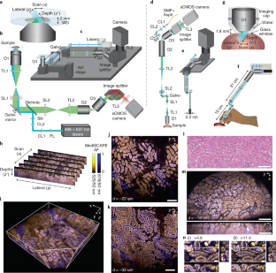
・MediSCAPEは、組織構造の画像を撮影することができる高速3D顕微鏡で、組織を切除して病理結果を待つ必要なく、外科医が腫瘍とその境界をナビゲートするための指針となることが期待されます。
・この研究の筆頭著者であるクリッパ・パテルは言う。「通常は目に見えないほど弱い、組織内の自然な蛍光を我々の顕微鏡は3Dボリューム全体をリアルタイムで歩き回れるほどのスピードでイメージングし、まるで懐中電灯を持っているかのように、これらの弱い信号をよく見ることが出来ました。」
<関連情報>
- https://www.engineering.columbia.edu/news/mediscape-3d-microscope-tissue-imaging
- https://www.nature.com/articles/s41551-022-00849-7
高速ライトシート顕微鏡による生体組織の体積組織像のin-situ取得 High-speed light-sheet microscopy for the in-situ acquisition of volumetric histological images of living tissue
Kripa B. Patel,Wenxuan Liang,Malte J. Casper,Venkatakaushik Voleti,Wenze Li,Alexis J. Yagielski,Hanzhi T. Zhao,Citlali Perez Campos,Grace Sooyeon Lee,Joyce M. Liu,Elizabeth Philipone,Angela J. Yoon,Kenneth P. Olive,Shana M. Coley &Elizabeth M. C. Hillman
Nature Biomedical Engineering (2022) Published: 28 March 2022

Abstract
Histological examinations typically require the excision of tissue, followed by its fixation, slicing, staining, mounting and imaging, with timeframes ranging from minutes to days. This process may remove functional tissue, may miss abnormalities through under-sampling, prevents rapid decision-making, and increases costs. Here, we report the feasibility of microscopes based on swept confocally aligned planar excitation technology for the volumetric histological imaging of intact living tissue in real time. The systems’ single-objective, light-sheet geometry and 3D imaging speeds enable roving image acquisition, which combined with 3D stitching permits the contiguous analysis of large tissue areas, as well as the dynamic assessment of tissue perfusion and function. Implemented in benchtop and miniaturized form factors, the microscopes also have high sensitivity, even for weak intrinsic fluorescence, allowing for the label-free imaging of diagnostically relevant histoarchitectural structures, as we show for pancreatic disease in living mice, for chronic kidney disease in fresh human kidney tissues, and for oral mucosa in a healthy volunteer. Miniaturized high-speed light-sheet microscopes for in-situ volumetric histological imaging may facilitate the point-of-care detection of diverse cellular-level biomarkers.


