2025-04-30 高輝度光科学研究センター,理化学研究所

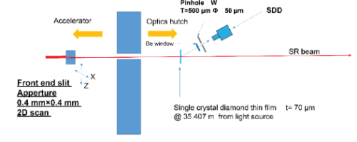
図1 広視野X線ビームモニターのセットアップ図。 図の左から放射光(SR beam)が入射し、FES(フロントエンドスリット)を通って、ダイヤモンド薄膜を通過する。散乱光はピンホールを通り、SDD(シリコンドリフト検出器)で検出される。
<関連情報>
- http://www.spring8.or.jp/ja/news_publications/press_release/2025/250430/
- https://journals.iucr.org/s/issues/2025/03/00/gy5074/index.html
ダイヤモンド薄膜とシリコンドリフト検出器を用いた放射光X線ビームの詳細プロファイルの可視化法
Method for visualizing detailed profiles of synchrotron X-ray beams using diamond-thin films and silicon drift detectors
Togo Kudo, Shinji Suzuki, Mutsumi Sano,Toshiro Itoga, Hiroyasu Masunaga,Shunji Goto and Sunao Takahashi
Journal of Synchrotron radiation Published:22 April 2025
DOI:https://doi.org/10.1107/S1600577525002838
Contamination from nearby bending magnet radiation hinders precise and accurate determination of the true beam center of undulator radiation. To solve this problem, a semi-nondestructive method was developed to visualize the detailed profile of a synchrotron radiation beam by using a thin diamond film as a scatterer. As the beam passed through the diamond film, scattered X-rays were imaged using a pinhole camera and measured with a two-dimensional silicon drift detector (SDD) scan. With this configuration, the beam center was accurately determined by visualizing the radiation pattern distribution for each energy level of a pink X-ray beam within an aperture size of 1.5 mm × 1.5 mm, shaped by a front-end slit (FES) positioned upstream of the monochromator. Additionally, by scanning the FES in two dimensions with a reduced aperture of 0.4 mm × 0.4 mm, energy-resolved images were successfully obtained using the SDD at a fixed position. These images revealed the profile of undulator radiation over a broad area (with an aperture extending up to 4 mm) in a pre-slit positioned upstream of the FES, demonstrating good alignment with SPECTRA calculations. This method effectively eliminates contamination from nearby bending magnet radiation, a significant issue in previous approaches, enabling a direct and highly accurate determination of the true beam center.


