2025-07-11 カリフォルニア大学アーバイン校(UCI)
<関連情報>
- https://news.uci.edu/2025/07/11/unraveling-molecular-secrets/
- https://www.nature.com/articles/s43586-025-00403-0
光誘起力顕微鏡 Photo-induced force microscopy
Maxim R. Shcherbakov,Eric O. Potma,Yasuhiro Sugawara,Derek Nowak,Mariia Stepanova,Philip R. Davies,Josh Davies-Jones & H. Kumar Wickramasinghe
Nature Reviews Methods Primers Published:29 May 2025
DOI:https://doi.org/10.1038/s43586-025-00403-0

Abstract
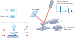
Photo-induced force microscopy (PiFM) has emerged as a transformative technique in nanoscale imaging, providing insights into the chemical composition and spatial organization of materials at the nanometre scale. PiFM enables innovation in surface science, geological research, biological studies, materials science, photonics and beyond. Compared with other probe-based spectroscopic methods, the nature of tip–sample interaction in PiFM can offer superior spatial resolution and surface sensitivity. This Primer offers the reader an introduction to the fundamentals of PiFM, discusses the configuration of the method, as well as advanced modalities and modifications, such as ultrahigh vacuum and nonlinear techniques. Results and scenarios are summarized, including those in surface chemistry, biology, polymer science and nanophotonics, offering a unique perspective for researchers across multiple disciplines. The Primer concludes with a discussion of PiFM’s limitations and points of improvement, and it outlines the future developments of this versatile nanoscale spectroscopy technique.


