2025-08-01 東北大学

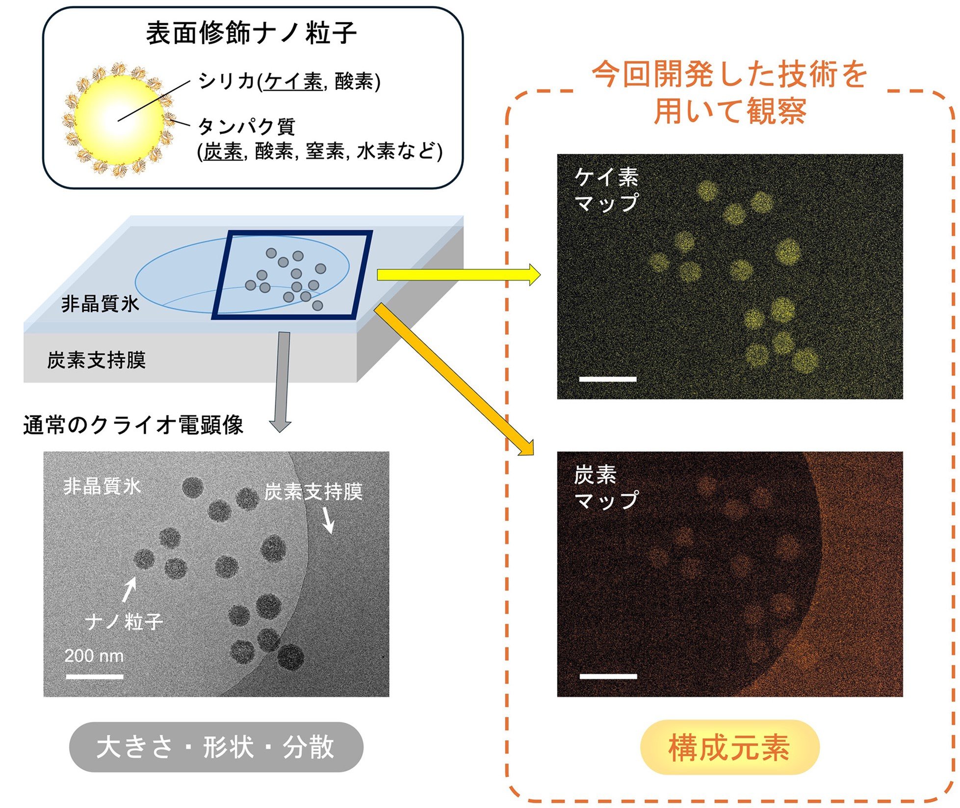
図1. クライオ電子顕微鏡を用いた表面修飾ナノ粒子の元素マッピング観察。(左下)通常のクライオ電顕像からは、ナノ粒子の大きさ・形状・分散状態などが分かる。(右上)ケイ素のマッピングでは、シリカナノ粒子由来のケイ素が検出・可視化された。(右下)炭素のマッピングでは、表面修飾タンパク質由来の炭素に加え、グリッド支持膜由来の炭素が検出・可視化された。
<関連情報>
- https://www.tohoku.ac.jp/japanese/2025/08/press20250801-03-nano.html
- https://www.tohoku.ac.jp/japanese/newimg/pressimg/tohokuuniv-press20250801_01web_nano.pdf
- https://pubs.acs.org/doi/10.1021/acs.analchem.5c02121
液体相材料中の軽元素の低線量元素マッピングをクライオ-EELSを用いて実施 Low-Dose Elemental Mapping of Light Atoms in Liquid-phase Materials Using Cryo-EELS
Daisuke Unabara,Yohei K. Sato,Tasuku Hamaguchi,and Koji Yonekura
Analytical Chemistry Published July 31, 2025
DOI:https://doi.org/10.1021/acs.analchem.5c02121
Abstract
Cryogenic transmission electron microscopy (cryo-TEM) enables the visualization of liquid-phase materials, including nanoparticles and soft/biomaterials, under cryogenic conditions while minimizing radiation damage. Cryo-TEM imaging provides insights into particle size, shape, and dispersion. Beyond such conventional structural information, acquiring elemental composition data allows for a more detailed analysis and evaluation. In this study, we developed a method for elemental mapping of nanoparticles and soft/biomaterials in frozen solvent by integrating electron energy loss spectroscopy (EELS) with energy-filtered (EF) cryo-EM. Cryo-EELS and analysis were performed using the three-window method, following the alignment and correction of stage drift during image acquisition and interexposure drift between different energy windows. This approach, by balancing accurate and reliable signal extraction with electron dose, enabled the generation of elemental maps for nanoparticles as small as 10 nm in frozen solvent. Furthermore, we extended the technique to protein-coated silica nanoparticles and hydroxyapatite (HAp) nanoparticles in vitrified solvent, selecting specific energy loss values to identify multiple constituent elements. As a result, we successfully mapped silica from the nanoparticle cores, carbon from the protein shell of the nanoparticles, and phosphorus and calcium─key light elements in biological systems─within the same imaging area of the HAp particles.


