2024-06-07 ニューヨーク大学 (NYU)
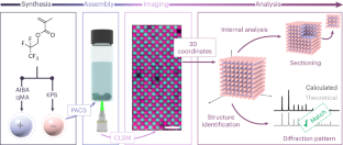
◆研究では、コロイド結晶を対象にしており、双晶現象や結晶が溶ける過程も観察できます。実験結果はコンピューターシミュレーションと一致し、結晶の成長過程を最適化するための重要な手がかりを提供します。これにより、フォトニック材料などの新素材の開発が期待されます。
<関連情報>
- https://www.nyu.edu/about/news-publications/news/2024/june/x-ray-vision-crystal-clear.html
- https://www.nature.com/articles/s41563-024-01917-w
イオン性コロイド結晶化の3次元実空間解析が可能に Enabling three-dimensional real-space analysis of ionic colloidal crystallization
Shihao Zang,Adam W. Hauser,Sanjib Paul,Glen M. Hocky & Stefano Sacanna
Nature Materials Published:03 June 2024
DOI:https://doi.org/10.1038/s41563-024-01917-w

Abstract
Structures of molecular crystals are identified using scattering techniques because we cannot see inside them. Micrometre-sized colloidal particles enable the real-time observation of crystallization with optical microscopy, but in practice this is still hampered by a lack of ‘X-ray vision’. Here we introduce a system of index-matched fluorescently labelled colloidal particles and demonstrate the robust formation of ionic crystals in aqueous solution, with structures that can be controlled by size ratio and salt concentration. Full three-dimensional coordinates of particles are distinguished through in situ confocal microscopy, and the crystal structures are identified via comparison of their simulated scattering pattern with known atomic arrangements. Finally, we leverage our ability to look inside colloidal crystals to observe the motion of defects and crystal melting in time and space and to reveal the origin of crystal twinning. Using this platform, the path to real-time analysis of ionic colloidal crystallization is now ‘crystal clear’.


