2024-09-27 オランダ・デルフト工科大学(TUDelft)
<関連情報>
- https://www.tudelft.nl/en/2024/tnw/first-ever-imaging-of-pathogens-on-lettuce-leaves-in-real-time
- https://www.nature.com/articles/s41467-024-52594-x
ラベルフリー光コヒーレンストモグラフィにより、植物の生体内病原体動態をリアルタイムで3D可視化 Revealing real-time 3D in vivo pathogen dynamics in plants by label-free optical coherence tomography
Jos de Wit,Sebastian Tonn,Mon-Ray Shao,Guido Van den Ackerveken & Jeroen Kalkman
Nature Communications Published:27 September 2024
DOI:https://doi.org/10.1038/s41467-024-52594-x

Abstract
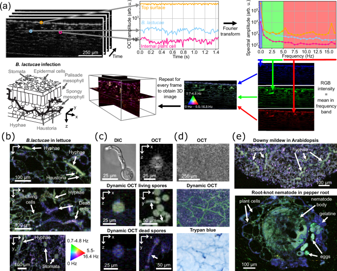
Microscopic imaging for studying plant-pathogen interactions is limited by its reliance on invasive histological techniques, like clearing and staining, or, for in vivo imaging, on complicated generation of transgenic pathogens. We present real-time 3D in vivo visualization of pathogen dynamics with label-free optical coherence tomography. Based on intrinsic signal fluctuations as tissue contrast we image filamentous pathogens and a nematode in vivo in 3D in plant tissue. We analyze 3D images of lettuce downy mildew infection (Bremia lactucae) to obtain hyphal volume and length in three different lettuce genotypes with different resistance levels showing the ability for precise (micro) phenotyping and quantification of the infection level. In addition, we demonstrate in vivo longitudinal imaging of the growth of individual pathogen (sub)structures with functional contrast on the pathogen micro-activity revealing pathogen vitality thereby opening a window on the underlying molecular processes.


